Magnified View (via Quicktime VR)
a) Frontal bone.
b) Greater wing of the sphenoid bone (s. superficial temporal bone).
c) Maxillary bone.
d) Medial wall of the orbit (lamina papyracea and os lacrymale).
e) Zygomatic bone.
f) Mandibular ramus.
g) Mandibular body.
h) m. External pterygoid.
i) m. Internal pterygoid.
k) m. Masseter.
l) m. Orbicularis oris.
m) m. Buccinator.
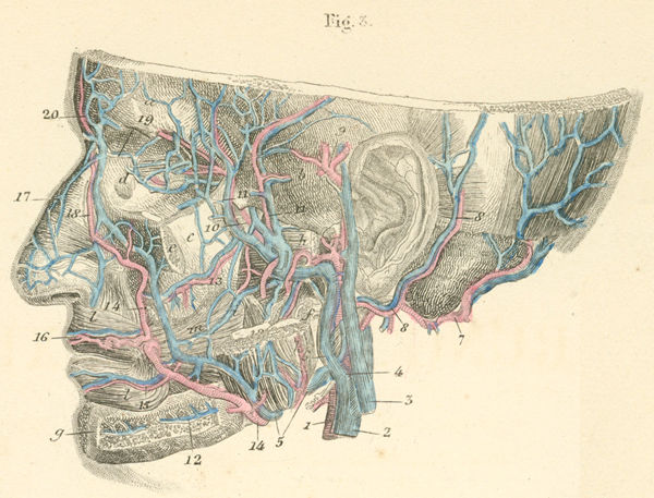
- left common carotid.
- internal jugular vein.
- external jugular vein.
- common facial vein (s. cephalica externa).
- facial vein.
- retromandibular vein.
- occipital artery and vein.
- posterior auricular vein.
- superficial temporal artery.
- maxillary artery.
- deep temporal artery.
- inferior alveolar artery and vein.
- posterior dental artery.
- facial artery (s. maxillaris externa).
- inferior labial artery (s. coronaria labii inferioris).
- superior labial artery (s.coronaria labii superioris).
- dorsal nasal artery.
- angular artery.
- ophthalmic artery and superior ophthalmic vein.
- frontal artery and vein.
Tidak ada komentar:
Posting Komentar